How do pregnancy ultrasounds work?
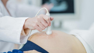
A probe is used to pass sound waves into the body. An image of the baby is produced by the reflection of those sound waves. The images (sonogram) are displayed on a television screen. The sonographer performing the ultrasound can explain these images to you. Appropriate use of ultrasound poses no physical risk to you or your baby.
Why would you have a prenatal ultrasound?
You may have an ultrasound to check the development of your pregnancy.
Confirming the stage of the pregnancy
Checking the continuation of a pregnancy if there has been bleeding; Identifying a multiple pregnancy
Checking the position of the placenta
Checking the amount of amniotic fluid
Checking the physical development of the foetus
Diagnosing birth defects and/or hereditary conditions.
A prenatal diagnostic ultrasound can detect physical abnormalities including:
Congenital heart defects : Abnormalities of the structure and function of the heart occur in 1 in 100 pregnancies
Limb reduction defects : Short or missing bones in the arm or leg occur in 1 in 1000 pregnancies
Cleft lip palate : Failure of normal closure of tissue in the lip palate area occurs in 1 in 1000 pregnancies
Neural tube defects : Abnormalities of the brain, skull, spine and spinal cord
How is an ultrasound performed?
There are two ways in which obstetric ultrasound can be performed.
Abdominal ultrasound
Abdominal ultrasound is the most common procedure and can be used at any stage of pregnancy
A gel is applied to the abdomen to allow the sound waves to pass from the transducer into the uterus
The bladder should be comfortably full to gain a clear ultrasound picture.
Vaginal ultrasound
Vaginal ultrasound is less commonly performed. It is usually restricted to the first trimester (up to 12 weeks) of pregnancy. The bladder needs to be completely emptied in order to perform this method of ultrasound.
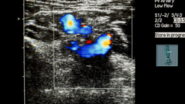
Doppler Scan in Pregnancy
Doppler sonography is a technique which uses reflected sound waves to measure movements such as blood flow and heartbeat. Doppler ultrasound scans can be used to determine the speed of the blood flow and its direction. This information can be helpful in determining if the foetal growth is normal and whether the tissues are supplied with enough blood and nutrients.Doppler scans are performed with the same apparatus as a regular ultrasound scan and are normally used during the third trimester on women who have high-risk pregnancies.
What Is Doppler Scan?
A Doppler scan is similar to a regular ultrasound scan and works using high-frequency sound waves called ultrasound that aren’t audible to our ears. The ultrasound generated by the equipment bounces off bones and tissues like an echo and is recorded with a microphone. All of this is done with a small hand-held probe called a transducer. A gel which helps in the process is applied over the belly, and the transducer is pressed gently against the skin to scan. Denser substances like bones give off a better echo than the softer tissue that the body is made up of, and by comparing the echo, an image of the baby is generated in a computer and displayed in real time.What sets a Doppler scan apart is that unlike a regular ultrasound scan, it can detect the flow of blood in blood vessels, estimate the speed of the blood flow, determine its direction, detect blood clots, etc. Most ultrasound equipment these days has an inbuilt Doppler feature and both the scans can be done together.